
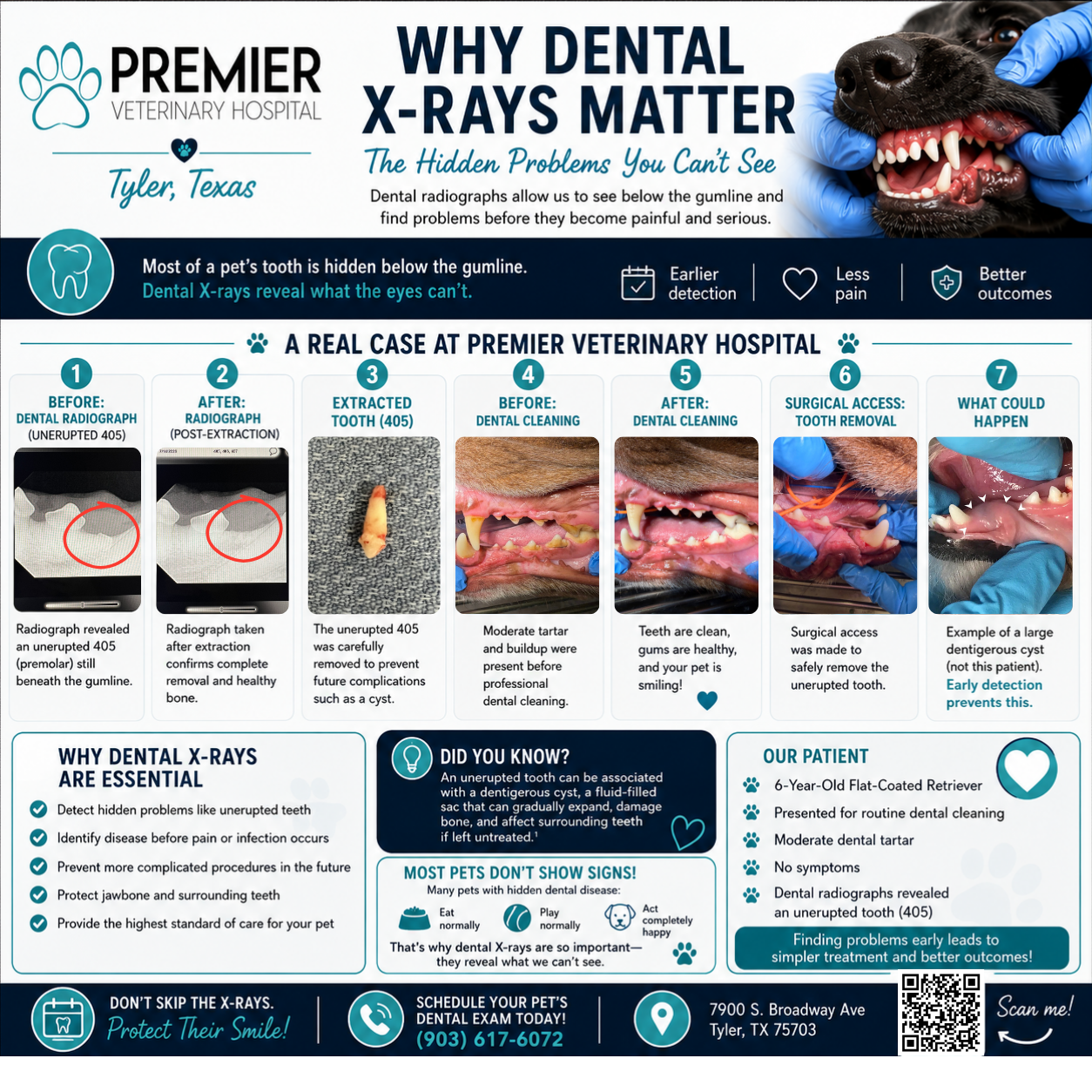
The Hidden Problems You Can’t See
A real case from Premier Veterinary Hospital: A 6-year-old Flat-Coated Retriever came in for a routine dental cleaning with moderate tartar and no obvious symptoms. Dental radiographs revealed an unerupted mandibular first premolar – tooth 405 – hidden beneath the gumline. The tooth was removed proactively to help prevent future complications.
This case shows why a dental cleaning is more than polishing the visible teeth. Radiographs help the veterinary team evaluate the roots, jawbone, and structures beneath the gumline – areas that cannot be assessed by sight alone.
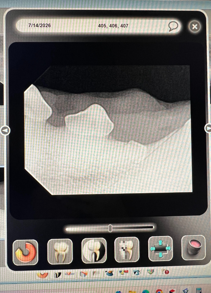
Pre-extraction dental radiograph showing the unerupted tooth
A Routine Visit Revealed a Hidden Problem
The patient presented for a professional dental cleaning because of moderate tartar. There was no facial swelling, difficulty eating, pawing at the mouth, or other outward sign that a tooth was hidden beneath the gumline.
Once the patient was safely anesthetized, the team completed a thorough oral examination and obtained full-mouth dental radiographs.
One image showed that tooth 405 had not erupted normally and remained embedded beneath the gum.
Without radiographs, this finding could easily have gone unnoticed. The mouth may look normal above the gumline while disease or developmental abnormalities are present below it.
Why this matters: When a tooth appears to be missing, radiographs help determine whether it is truly absent, previously extracted, fractured, or unerupted.
Why Dental Radiographs Are Essential
A large portion of every tooth lies below the gumline. Dental radiographs allow veterinarians to evaluate tooth roots, surrounding bone, retained roots, fractures, abscesses, tooth resorption, impacted teeth, and developmental abnormalities. They also help the doctor plan treatment and confirm that a tooth and its roots have been completely removed.
The educational review from Today’s Veterinary Nurse explains that unerupted teeth can be associated with dentigerous cyst formation. These cysts are often found incidentally because many patients show no discomfort or swelling when the lesion is still small.
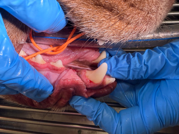
Surgical access to the unerupted tooth (#405).
Treatment: Removing Tooth 405 Before Complications Developed
Because tooth 405 remained unerupted, Dr. Hogue recommended surgical removal. The goal was preventive: remove the tooth before it could contribute to future problems, including cyst formation, bone damage, or injury to nearby teeth.
The gum tissue was carefully opened to access the unerupted tooth. After extraction, a postoperative radiograph was obtained to confirm complete removal and evaluate the surrounding bone.
No dentigerous cyst was confirmed in this patient. The tooth was removed because of the risk associated with leaving an unerupted tooth in place.
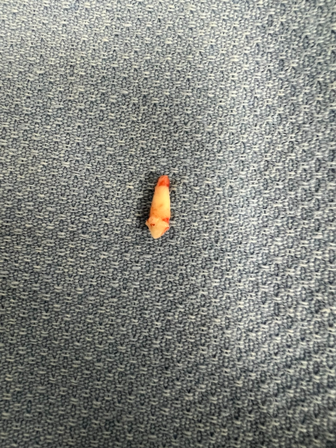
Extracted tooth 405
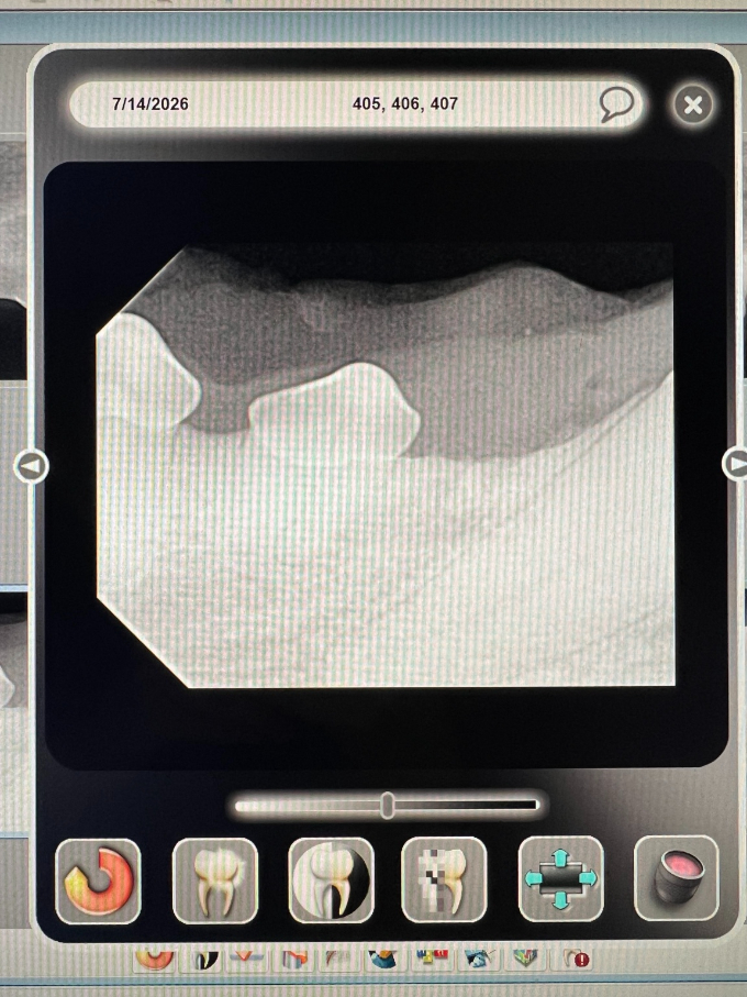
Post-extraction radiograph confirming removal.
Did you know?
A dentigerous cyst is a fluid-filled structure associated with an unerupted tooth. Over time, it may expand and damage bone or nearby teeth. Early detection usually means a simpler procedure and a better outcome.
Before and After the Dental Cleaning
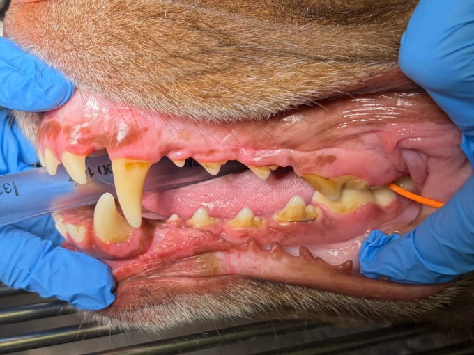
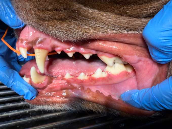
Before: Moderate tartar and buildup.
After: Clean teeth and healthier-looking gums.
A professional dental cleaning removes plaque and tartar from above and below the gumline. But the radiographs are what allowed the team to identify the hidden tooth and make a complete treatment plan. The cleaning improved the visible health of the mouth, while the radiographs helped address what could not be seen.
What Could Happen Without Early Detection?
The image at right is an educational example from the Today’s Veterinary Nurse article and is not this patient. It shows the gross appearance of a large dentigerous cyst with gray fluid visible through the oral mucosa.
This example demonstrates why apparently missing teeth should not be ignored. Annual oral examinations, professional dental cleanings when recommended, and dental radiographs can help identify hidden problems before they become extensive.
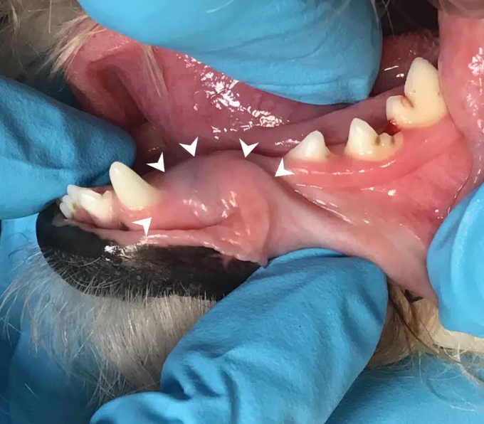
Educational example of a large dentigerous cyst. Image credit: Sheena Davis, LVT, VTS (Dentistry), Today’s Veterinary Nurse, Winter 2024.
Most Pets Do Not Show Obvious Signs
| Pets with hidden dental disease may still: | Dental radiographs can help identify: |
|---|---|
|
|
The Take-Home Message
This patient came in for a routine dental cleaning and showed no signs of pain, yet radiographs revealed an unerupted tooth that required treatment. Finding it early allowed the veterinary team to remove the tooth before a cyst or other complication was confirmed.
Comprehensive dental care includes an anesthetized oral examination, professional cleaning, and full-mouth dental radiographs. Together, these steps allow the veterinary team to care for both the visible crown of each tooth and the hidden structures below the gumline.
Protect your pet’s smile.
Schedule a dental evaluation with Premier Veterinary Hospital.
(903) 617-6072 | 7900 S. Broadway Ave, Tyler, TX 75703
Reference: Davis S. Dentigerous Cysts: An Overview. Today’s Veterinary Nurse. Winter 2024;76-81.